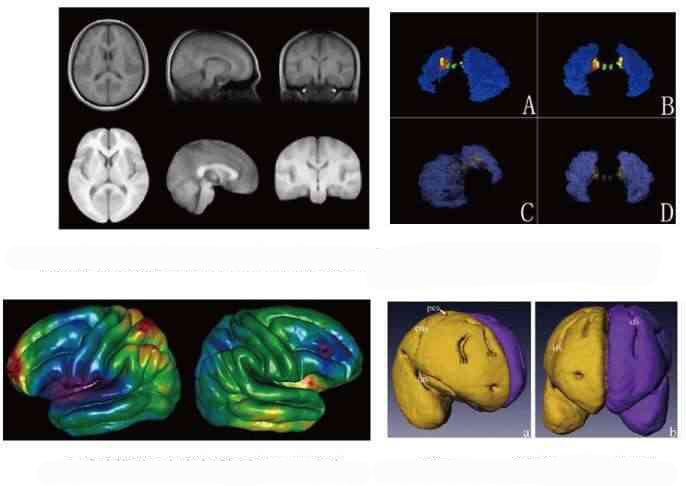
Establishment of the human brain atlas and its variation
Using in vivo and specimen CT and MRI, to construct interactive digital brain atlas and study the race, age, hemisphere and gender differences. With the help of Archaeology, this atlas can be a powerful tool to support the research of neuroscience and digital medicine. Our researches were published in NeuroImage, Brain Research and Journal of Anatomy.
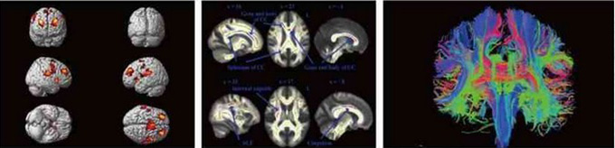
Functional neuroimaging
Using fMRI and DTI to study brain functions in vivo. Nowadays, our group used three-dimensional MRI image of the twins brain and studied the neural basis and degree of genetic of attention and attention deficit. Our researches were published on Human Brain Mapping and Acta Radiologica.
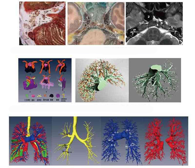
Digital surgical anatomy
NC frozen milling technology can obtain the human specimen cross-section in sub-millimeter. With the help of computer virtual reality technology, we can construct three dimensional visualization and virtualization of the human brain formation, which can provides the basis for the development of minimally invasive surgery and digital surgery. Our group focus on the study of digital anatomy of the skull, brain, liver, and lung. Our researches were published in World Neurosurgery, Surgical and Radiologic Anatomy.
Molecular imaging study
Using ex vivo and in vitro PD models and molecular imaging of cellular and molecular biology techniques to study the mechanism the pathogenesis and treatment of Parkinson's disease. Our researches were published in Brain Research and J Neurosci Res.
